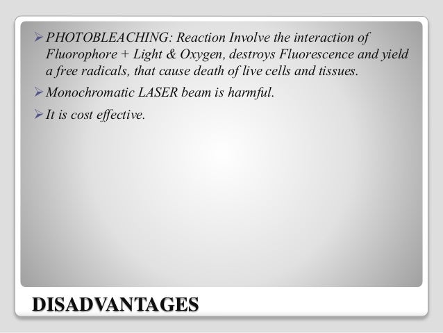
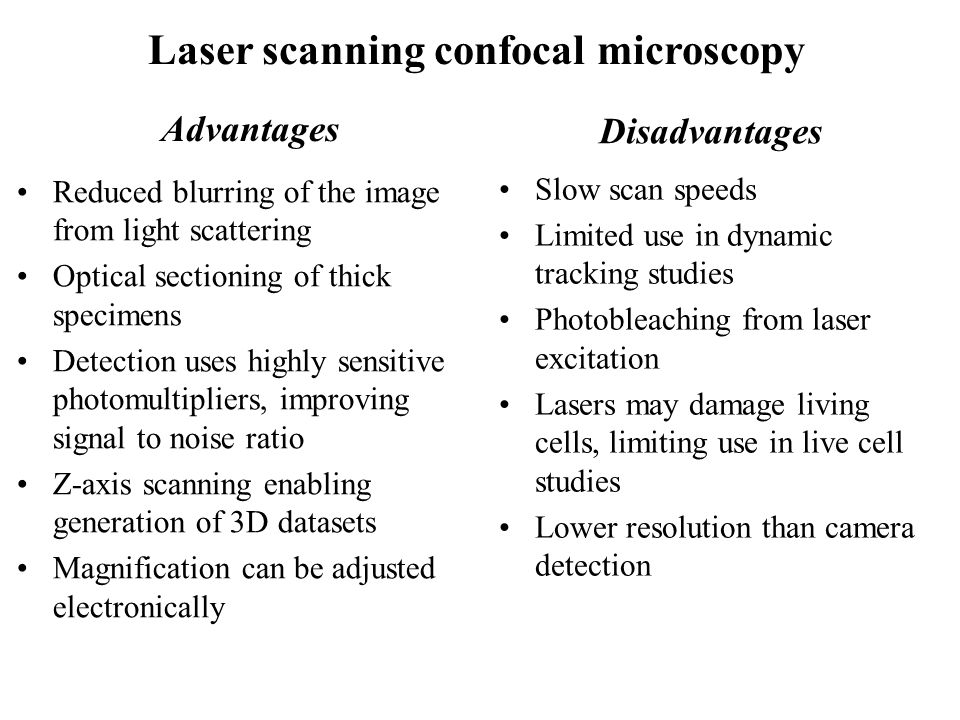
Disadvantages Of Laser Scanning Microscopes

What Are The Limitations Of Confocal Laser Scanning Microscopes Quora
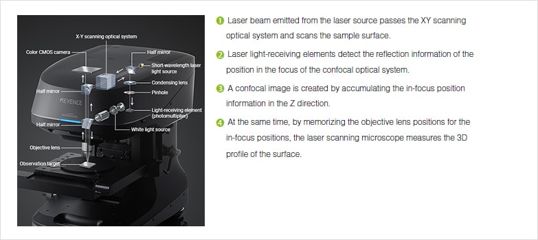
Profile Measuring Laser Microscopes Instruments Used For Roughness Measurements Introduction To Roughness Keyence America

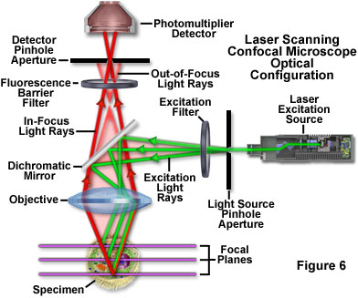
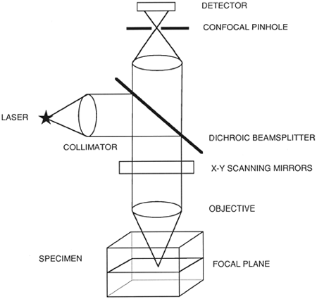
What Is Confocal Laser Scanning Microscopy

Modern Laser Scanning Confocal Microscopy Bayguinov 2018 Current Protocols In Cytometry Wiley Online Library

Tevo Tornado X Axis Tensioner Remix By Gabix Thingiverse Tornado Remix 3d Printing

If You Need Any Optical And Ophthalmic Device Pls Contact Me Email Lisafan Hyvisionstar Com Eye Health Ophthalmic Equipment Optical
Advantages and disadvantages of confocal microscopy.
Disadvantages of laser scanning microscopes.
Confocal Laser Scanning Microscopy Clsm

Orlas Station Machine Design Cnc Design Industrial Machine
Confocal Microscopy Introduction Olympus Life Science

The Use Of Laser Scanning Confocal Microscopy Lscm In Materials Science Hovis 2010 Journal Of Microscopy Wiley Online Library

Tattooremovalproducts Laser Hair Removal Machine Hair Removal Machine Tattoo Removal

Manual Capsulorhexes Above And Catalys Capsulotomies Below Stained With Trypan Blue Catalys Capsulotomies Exhibit Precisio Cataract Surgery Cataract Laser
Zeiss Microscopy Online Campus Live Cell Imaging Microscopy Techniques
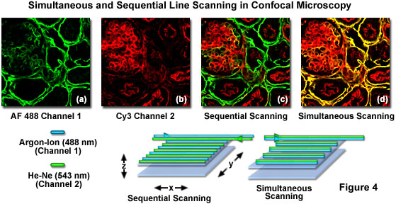
Olympus Fluoview Resource Center Spectral Bleed Through Artifacts In Confocal Microscopy

World S First White Lasers Demonstrated More Luminous Energy Efficient Than Leds White Lasers Look To Be The Future In Lighting And Li Fi Or Light Based Wir Futuristic Technology Nanotechnology Technology

Confocal Microscopy Comparisons Applications And Problems Biotechniques

What Is Spinning Disk Confocal Microscopy
Confocal Laser Scanning Microscopy Springerlink

Laser Microscope An Overview Sciencedirect Topics

Researchers Develop First Ever Single Molecule Led First Ever Science Oxford Brookes University

Digital Podium Is The State Of The Art Solution For Smart Conference Room Smart Classroom And Auditorium I Interactive Presentation Digital Activities Podium
Http Www Microscopist Co Uk Wp Content Uploads 2017 04 Artefacts In Confocal Pdf
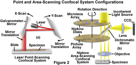
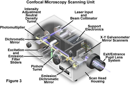
Confocal Microscopy Confocal Microscope Scanning Systems Olympus Life Science
Fluorescence And Confocal Microscopy Ppt Video Online Download
Https Encrypted Tbn0 Gstatic Com Images Q Tbn 3aand9gcr3fysoxor5w4y0kayjtt5nby84 Yhi3vdxn3rx2 E Usqp Cau
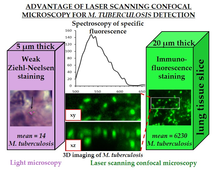
Jcm Free Full Text Application Of Laser Scanning Confocal Microscopy For The Visualization Of M Tuberculosis In Lung Tissue Samples With Weak Ziehl Neelsen Staining Html

Focal Wars Widefield Vs Confocal Biocompare The Buyer S Guide For Life Scientists

Attendance In Excel Sheet Using Rfid Rc522 Hackster Io Arduino Rfid Arduino Beginner

Confocal Laser Scanning Microscopy An Overview Sciencedirect Topics
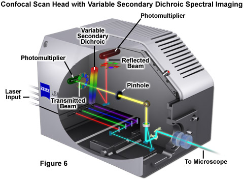
Zeiss Microscopy Online Campus Introduction To Spectral Imaging
Source : pinterest.com
